Dr. R, arguably the best equine lameness diagnostician in Vermont, came out on February 5th to take images of both hocks and ultrasound the RH, which was the original injured leg. Stella had been at the new barn for a couple weeks by then, and was settling nicely into "doing stuff" that resembled rehab. She had been chiropracted by Dr. R the week before this visit, but something in my gut told me I really needed to see what exactly was going on in her joints, if not just to confirm that she was healed and ready for a job.
Glad I did.

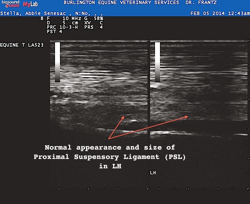

The difference between the RH and LH suspensories is HUGE when you look at them together. For those who aren't savvy at reading u/s images, the first image is take looking at the back of her leg, u/s a few inches below the point of the hock. In the second and third, the images are flipped so the horse's leg is viewed as if it were horizontal.
I think the actual measurement difference between the two suspensories was almost 4mm. The LH measured something like 9.3mm, and if her RH had matched up approximately, there'd be no concern. But 4mm is a big difference, which indicates some kind of trauma. You can also see, in the first image, that it's enlarged: you also get the benefit there of seeing all the fuzzy grey, which is irregular scar tissue patterns, indicating it was injured but is now healed.
Yes, good news is, healed. Also good news, no lesions showed up in the u/s, which would make her chances of re-injury much higher.
Coming up: x-rays
No comments:
Post a Comment